Tom Goddard
November 17, 2008
Representation of viruses in the remediated PDB archive.
Lawson CL, Dutta S, Westbrook JD, Henrick K, Berman HM.
Acta Crystallogr D Biol Crystallogr. 2008 Aug;D64(Pt 8):874-82. Epub 2008 Jul 17.
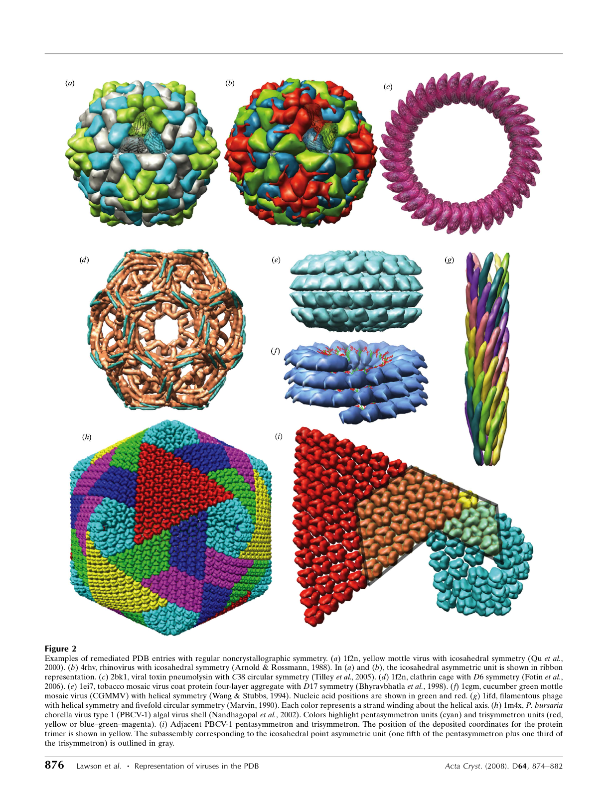
Paramecium bursaria chorella virus type 1 (PBCV-1) is PDB entry 1m4x.
Has 3 chains A,B,C forming a trimer.
1680 trimers for the capsid.
#
loop_
_pdbx_struct_assembly.id
_pdbx_struct_assembly.details
1   'complete icosahedral assembly'
2   'icosahedral asymmetric unit'
3   'icosahedral pentamer'
4   'icosahedral 23 hexamer'
5   'pentasymmetron capsid unit'
6   'trisymmetron capsid unit'
PAU 'icosahedral asymmetric unit, std point frame'

#
loop_
_pdbx_struct_assembly_gen.assembly_id
_pdbx_struct_assembly_gen.oper_expression
_pdbx_struct_assembly_gen.asym_id_list
_pdbx_struct_assembly_gen.entity_inst_id
1   (1-60)(61-88)           A,B,C .
2   (61-88)                 A,B,C .
3   (1-5)(61-88)            A,B,C .
4   (1,2,6,10,23,24)(61-88) A,B,C .
5   (1-5)(63-68)            A,B,C .
6   (1,10,23)(61,62,69-88)  A,B,C .
PAU (P)(61-88)              A,B,C .

#
loop_
_pdbx_struct_oper_list.id
_pdbx_struct_oper_list.type
_pdbx_struct_oper_list.matrix[1][1]
_pdbx_struct_oper_list.matrix[1][2]
_pdbx_struct_oper_list.matrix[1][3]
_pdbx_struct_oper_list.vector[1]
_pdbx_struct_oper_list.matrix[2][1]
_pdbx_struct_oper_list.matrix[2][2]
_pdbx_struct_oper_list.matrix[2][3]
_pdbx_struct_oper_list.vector[2]
_pdbx_struct_oper_list.matrix[3][1]
_pdbx_struct_oper_list.matrix[3][2]
_pdbx_struct_oper_list.matrix[3][3]
_pdbx_struct_oper_list.vector[3]
P  'transform to point frame'    0.30901699  -0.50000000 0.80901699  0.00000  0.80901699  -0.30901699 -0.50000000 0.00000
0.50000000  0.80901699  0.30901699  -0.00000
1  'point symmetry operation'    1.00000000  0.00000000  0.00000000  0.00000  0.00000000  1.00000000  0.00000000  0.00000
0.00000000  0.00000000  1.00000000  0.00000
2  'point symmetry operation'    0.80901699  0.30901699  0.50000000  0.00000  0.30901699  0.50000000  -0.80901699 0.00000
-0.50000000 0.80901699  0.30901699  0.00000

...

60 'point symmetry operation'    -0.50000000 -0.80901699 -0.30901699 0.00000  0.80901699  -0.30901699 -0.50000000 0.00000
0.30901699  -0.50000000 0.80901699  0.00000
61 'build point asymmetric unit' 1.00000     0.00000     0.00000     0.0      0.00000     1.00000     0.00000     0.0      0.00000
0.00000     1.00000     0.0
62 'build point asymmetric unit' 0.999773    0.014247    -0.015805   -49.486  -0.014842   0.999161    -0.038166   14.993
0.015248    0.038393    0.999147    32.902

...

88 'build point asymmetric unit' 0.967112    0.161474    -0.196522   -130.071 -0.188316   0.973929    -0.126494   95.792
0.170973    0.159342    0.972305    -1.525


#
loop_
_pdbx_struct_legacy_oper_list.id
_pdbx_struct_legacy_oper_list.name
_pdbx_struct_legacy_oper_list.matrix[1][1]
_pdbx_struct_legacy_oper_list.matrix[1][2]
_pdbx_struct_legacy_oper_list.matrix[1][3]
_pdbx_struct_legacy_oper_list.vector[1]
_pdbx_struct_legacy_oper_list.matrix[2][1]
_pdbx_struct_legacy_oper_list.matrix[2][2]
_pdbx_struct_legacy_oper_list.matrix[2][3]
_pdbx_struct_legacy_oper_list.vector[2]
_pdbx_struct_legacy_oper_list.matrix[3][1]
_pdbx_struct_legacy_oper_list.matrix[3][2]
_pdbx_struct_legacy_oper_list.matrix[3][3]
_pdbx_struct_legacy_oper_list.vector[3]
1    1x61  1.00000000  0.00000000  0.00000000  0.00000    0.00000000  1.00000000  0.00000000  0.00000    0.00000000  0.00000000
1.00000000  0.00000
2    1x62  0.99977300  0.01424700  -0.01580500 -49.48600  -0.01484200 0.99916100  -0.03816600 14.99300   0.01524800  0.03839300
0.99914700  32.90200

...

1679 60x87 -0.28149639 -0.95353767 0.10734706  -20.06292  0.76749273  -0.29088592 -0.57126126 -188.26516 0.57594612  -0.07842024
0.81371768  -71.92725
1680 60x88 -0.38403872 -0.91790149 -0.09986197 -11.99060  0.75511638  -0.24999640 -0.60605334 -134.06850 0.53133210  -0.30815591
0.78912963  -89.32390
REMARK 300
REMARK 300 BIOMOLECULE: 1
REMARK 300 THIS ENTRY CONTAINS THE UNIQUE NON-CRYSTALLOGRAPHIC REPEAT
REMARK 300 UNIT, WHICH CONSISTS OF 3  CHAIN(S). SEE REMARK 350 FOR
REMARK 300 INFORMATION ON GENERATING THE BIOLOGICAL MOLECULE(S).
REMARK 300 THE ASSEMBLY REPRESENTED IN THIS ENTRY HAS REGULAR
REMARK 300 ICOSAHEDRAL POINT SYMMETRY (SCHOENFLIES SYMBOL = I).
REMARK 350
REMARK 350 GENERATING THE BIOMOLECULE
REMARK 350 COORDINATES FOR A COMPLETE MULTIMER REPRESENTING THE KNOWN
REMARK 350 BIOLOGICALLY SIGNIFICANT OLIGOMERIZATION STATE OF THE
REMARK 350 MOLECULE CAN BE GENERATED BY APPLYING BIOMT TRANSFORMATIONS
REMARK 350 GIVEN BELOW.  BOTH NON-CRYSTALLOGRAPHIC AND
REMARK 350 CRYSTALLOGRAPHIC OPERATIONS ARE GIVEN.
REMARK 350
REMARK 350 BIOMOLECULE: 1
REMARK 350 APPLY THE FOLLOWING TO CHAINS: A, B, C
REMARK 350   BIOMT1   1  1.000000  0.000000  0.000000        0.00000
REMARK 350   BIOMT2   1  0.000000  1.000000  0.000000        0.00000
REMARK 350   BIOMT3   1  0.000000  0.000000  1.000000        0.00000
REMARK 350   BIOMT1   2  0.999773  0.014247 -0.015805      -49.48600
REMARK 350   BIOMT2   2 -0.014842  0.999161 -0.038166       14.99300
REMARK 350   BIOMT3   2  0.015248  0.038393  0.999147       32.90200
...
REMARK 350   BIOMT1  1680 -0.384039 -0.917901 -0.099862      -11.99060
REMARK 350   BIOMT2  1680  0.755116 -0.249996 -0.606053     -134.06850
REMARK 350   BIOMT3  1680  0.531332 -0.308156  0.789130      -89.32390
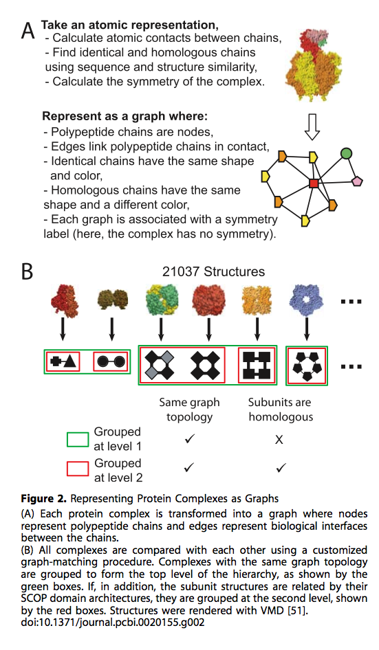
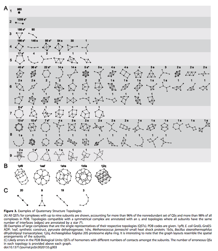
3D complex: a structural classification of protein complexes.
Levy ED, Pereira-Leal JB, Chothia C, Teichmann SA.
PLoS Comput Biol. 2006 Nov 17;2(11):e155. Epub 2006 Oct 5.
More than half of RCSB structures are homo- or heteromeric protein complexes.
Chimera does not recognize and identify identical or similar chains in multi-chain complexes.
Listing all unique contact interfaces between chains would be useful.
As of Nov 12 2008 there are 52444 entries loaded with the following exceptions:
nucleic acid and nuclei acid/protein complexes excluded all virus coat assemblies related entries excluded.
1bbs 1cde 1cyc 1hrb 1lbt 1xys 2cha 2lgs 1bcf
1gto 1xim 2xim 3pcn 3pca 3pcf 3pch 3pci 3pcj
3pck 3pcl 3pcm 3xim 1bcf 2lgs 123d 1ruo 1wio
1wip 3tra
The above entries are available via the PDBidcode search mechanism.
---------------------------------------------------------
 Oligomer         Number      Number       Number       Examples
  size            generated   homo-        hetero-
                   (accepted)   (accepted)    (accepted)
---------------------------------------------------------
 monomer/complex   19491
 dimer             17903 (16472)  13705 (12363)   4194  (4109)
 trimer             3554  (3496)   1664  (1611)   1890  (1885)
 tetramer           6013  (5962)   3823  (3785)   2190  (2177)
 pentamer            398   (394)    195   (195)    203   (199)   1BOV  (homo) 1A2K  (het)
 hexamer            1862  (1845)   1158  (1143)    704   (702)   1AEI  (homo) 1AFV  (het)
 heptamer            100    (99)     48    (48)     52    (51)   7AHL  (homo) 1LTI  (het)
 octamer             786   (779)    453   (450)    333   (329)   1AHU  (homo) 1BFV  (het)
 nonamer              88    (88)     11    (11)     77    (77)   1FWA
 decamer             157   (157)     91    (91)     66    (66)   1GTP
 undecamer            17    (17)     14    (14)      3     (3)   1WAP
 dodecamer           472   (472)    215   (215)    257   (257)   1COA  (homo) 1IZB  (het)
 tetradecamer         46    (46)     34    (34)     12    (12)   1GRL  (homo) 1AVO  (het)
 hexadecamer          98    (98)     16    (16)     82    (82)   2HLC  (homo) 1AA1  (het)
 octadecamer          27    (26)      6     (6)     21    (20)   1KZU
 21meric              41    (41)      0     (0)     41    (41)   1AON  (homo)
 24meric             144   (140)     85    (85)     59    (55)   1AEW  (homo) 3PCA  (het)
 26meric              14    (14)      0     (0)     14    (14)   1OCC
 28meric              21    (21)      1     (1)     20    (20)   1PMA
---------------------------------------------------------
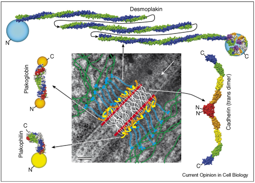
Desmosomes from a structural perspective.
Stokes DL.
Curr Opin Cell Biol. 2007 Oct;19(5):565-71. Epub 2007 Oct 22. Review.
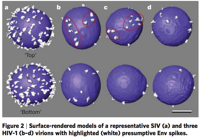
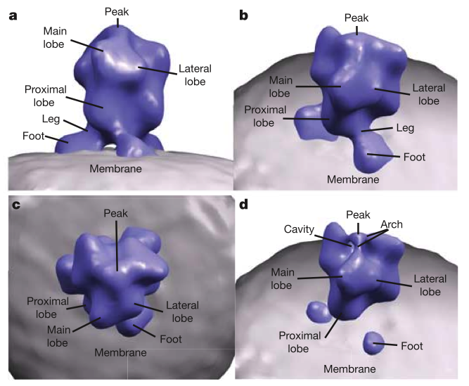
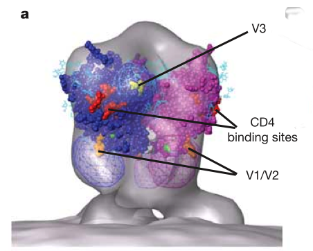
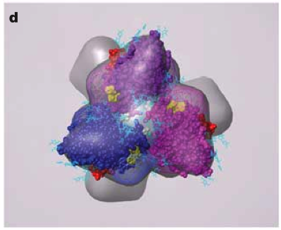
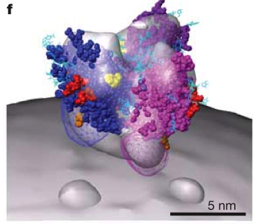
June 2006 EM tomography example fitting HIV gp120/gp41 spikes using Chimera. Spikes recognize target cells and initiate viral fusion. Illustrates combined use of tomography and structure averaging approach.
Zhu P, Liu J, Bess J Jr, Chertova E, Lifson JD, Grise H, Ofek GA, Taylor KA, Roux KH.
Distribution and three-dimensional structure of AIDS virus envelope spikes.
Nature. 2006 Jun 15;441(7095):847-52. Epub 2006 May 24.
|
|
|
| 
| 
|
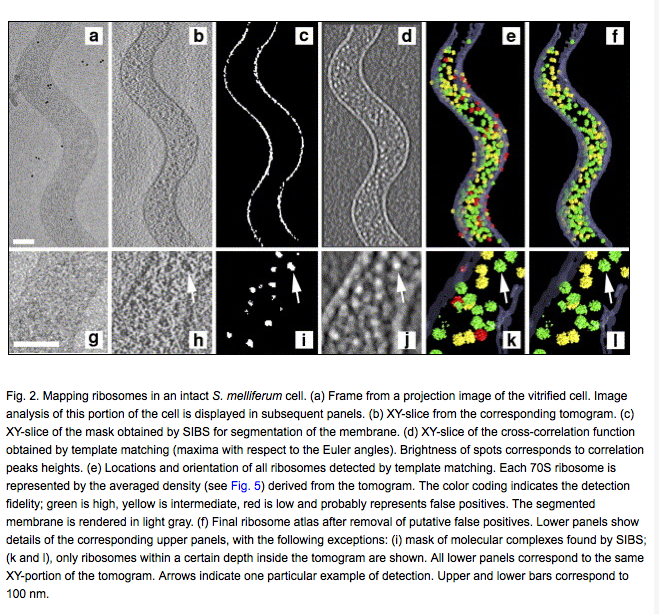
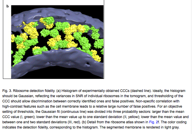
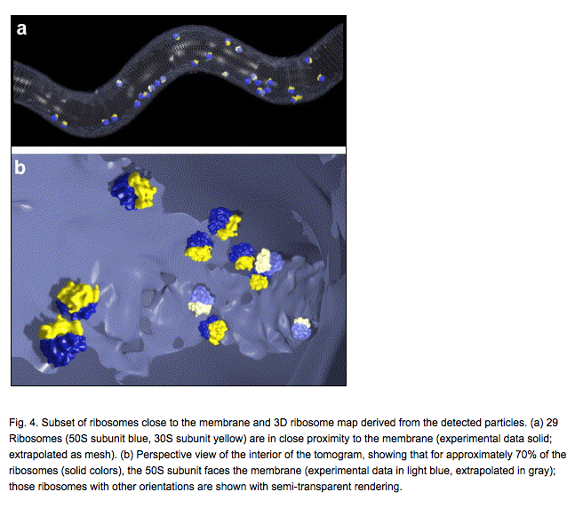
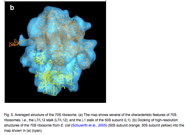
Mapping 70S ribosomes in intact cells by cryoelectron tomography and pattern recognition.
Ortiz JO, Förster F, Kürner J, Linaroudis AA, Baumeister W.
J Struct Biol. 2006 Nov;156(2):334-41. Epub 2006 Jun 3