
|
|
|
Depth cue
 JPEG version (118KB),
TIFF version (684KB)
JPEG version (118KB),
TIFF version (684KB)
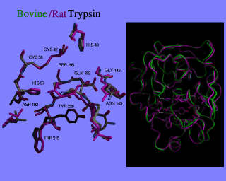
This image compares cow and rat trypsin. Depth cueing in neon was used to show the depth of the active site pocket, even though only a select number of active site residues are shown.
The image on the left was made using neon with a depth cue intensity by changing the following line in the neon.dat file being used (do a "man neon" for more information).
1 0.10 |Depthcue,Fraction |0=n, 1=y; percent aft intensityThis makes the "aft intensity" a tenth of the foreground, causing the tyrosine inside the pocket to appear darker and farther away, without having to have a stereo image. This allows for the overall comparison of the two proteins to be shown on the right, also using neon.
The image on the right, on the black background, was created separately, and scaled to be the right size with the SGI utility program "izoom". It was then pasted onto the full image of the active site, where the coordinates were displayed on the left hand side of the screen, using the SGI utility program "imgview -n filename".
The entire screen was then saved using the "scrsave" command. Do a "man" on any of these commands for more information:
- izoom
- imgview
- scrsave
- neon
Also see the description for the image two views for a description of cutting and pasting images together.
Image created by Julie Newdoll for Robert M. Stroud. Bovine Trypsin by R.M. Stroud, L.M. Kay and R.E. Dickerson. Rat Trypsin by R.M. Stroud and J. Finer-Moore. ©2004 The Regents, University of California; all rights reserved.