
|
|
|
Label 3D
 JPEG version
(67KB), TIFF version (296KB)
JPEG version
(67KB), TIFF version (296KB)
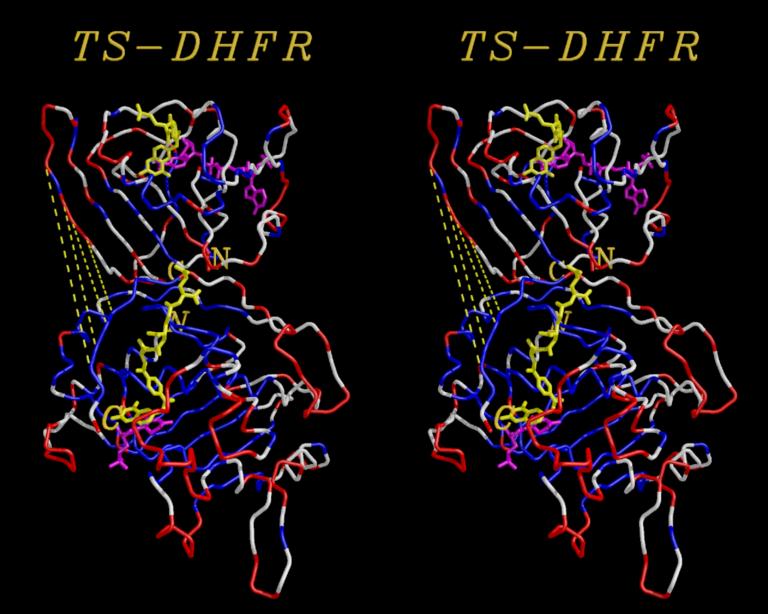
Using the MidasPlus delegates Neon, Label3D and Stereoimg, shown is a cross-eye stereo image of a monomer of E.coli Thymidylate Synthase (lower, TS) and E.coli Dihydrofolate Reductase (upper, DHFR) arranged roughly as determined for TS-DHFR complex (Knighton et. al., Nature Str1994),
The C and N termini of DHFR are labelled in bold, while the C and N termini of TS are labelled in bold italics. Both are colored according to electrostatic potential, where red represents electrostatic values less than -1, white values between -1 and +1, and blue are values greater than +1.
Yellow lines show the pathway along which the DHFR may hinge-bend onto the TS to bring the two protein active sites into close proximity (a theoretical mechanism).
See the manual pages on label3d, stereoimg and neon.
R.M. Stroud, Nature Structural Biology, May 1994. ©2004 The Regents, University of California; all rights reserved.