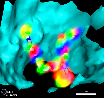
Light microscope volume showing fluorescently labeled Drosophila chromosome 2L. Three fluorophors label specific segments of the chromosome. The labeled segments are approximately 10 kilo-basepairs (kbp) in length and are separated by about 100 kbp. Segments of two homologous chromosomes are seen. The cyan surface shows the nuclear envelope determined from DAPI staining. The tubes were created with the Chimera pathtracer extension. The data is the unpublished work of Mike Lowenstein.
© 2004 The Regents of the University of California; all rights reserved.