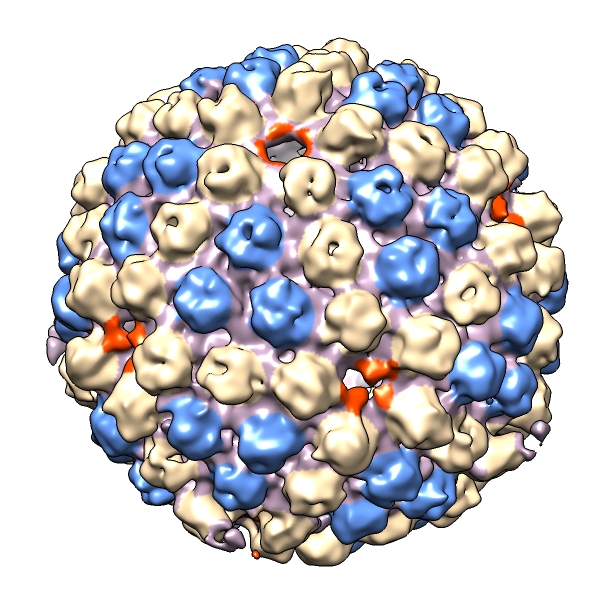
The arrangement of subunits in the capsid of herpes simplex virus type 1 is shown. Herpes virus has a portal at one of the icosahedron 5-fold vertices shown near the top in orange. The double-stranded DNA virus genome is packaged into the capsid via this portal, and when infecting a host cell is ejected through the portal and through a cell a nuclear pore.
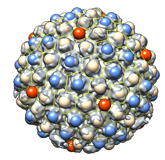
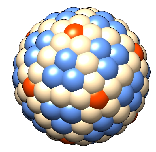
The coloring of the density map contour surface was done with the Chimera color zone tool. A marker was centered on each capsomere using the hkcage command (lattice parameters h = 4, k = 0) and a Python script markhidden.py that created the markers at the hidden lattice vertices (centers of hexagons and pentagons).
| 
|
The herpes density map shown is EMD-1307 from the Electron Microscopy Data Bank. It is a 5-fold averaged map about the portal vertex.
© 2009 The Regents of the University of California; all rights reserved.